What is Hip Dysplasia?
Hip Dysplasia is a terrible genetic disease because of the various degrees of arthritis (also called degenerative joint disease, arthrosis, osteoarthrosis) it can eventually produce, leading to pain and debilitation.

The very first step in the development of arthritis is articular cartilage (the type of cartilage lining the joint) damage due to the inherited bad biomechanics of an abnormally developed hip joint. Traumatic articular fracture through the joint surface is another way cartilage is damaged. With cartilage damage, lots of degradative enzymes are released into the joint. These enzymes degrade and decrease the synthesis of important constituent molecules that form hyaline cartilage called proteoglycans. This causes the cartilage to lose its thickness and elasticity, which are important in absorbing mechanical loads placed across the joint during movement. Eventually, more debris and enzymes spill into the joint fluid and destroy molecules called glycosaminoglycan and hyaluronate which are important precursors that form the cartilage proteoglycans. The joint's lubrication and ability to block inflammatory cells are lost and the debris-tainted joint fluid loses its ability to properly nourish the cartilage through impairment of nutrient-waste exchange across the joint cartilage cells. The damage then spreads to the synovial membrane lining the joint capsule and more degradative enzymes and inflammatory cells stream into the joint. Full thickness loss of cartilage allows the synovial fluid to contact nerve endings in the subchondral bone, resulting in pain. In an attempt to stabilize the joint to decrease the pain, the animal's body produces new bone at the edges of the joint surface, joint capsule, ligament and muscle attachments (bone spurs). The joint capsule also eventually thickens and the joint's range of motion decreases.
No one can predict when or even if a dysplastic dog will start showing clinical signs of lameness due to pain. There are multiple environmental factors such as caloric intake, level of exercise, and weather that can affect the severity of clinical signs and phenotypic expression (radiographic changes). There is no rhyme or reason to the severity of radiographic changes correlated with the clinical findings. There are a number of dysplastic dogs with severe arthritis that run, jump, and play as if nothing is wrong and some dogs with barely any arthritic radiographic changes that are severely lame.
An Examination of Hip Grading
The phenotypic evaluation of hips done by the Orthopedic Foundation for Animals falls into seven different categories. Those categories are Normal (Excellent, Good, Fair), Borderline, and Dysplastic (Mild, Moderate, Severe). Once each of the radiologists classifies the hip into one of the 7 phenotypes above, the final hip grade is decided by a consensus of the 3 independent outside evaluations. Examples would be:
- Two radiologists reported Excellent, one Good—the final grade would be Excellent
- One radiologist reported Excellent, one Good, one Fair—the final grade would be Good
- One radiologist reported Fair, two radiologists reported Mild—the final grade would be Mild
The hip grades of Excellent, Good and Fair are within normal limits and are given OFA numbers. This information is accepted by AKC on dogs with permanent identification (tattoo, microchip) and is in the public domain. Radiographs of Borderline, Mild, Moderate and Severely dysplastic hip grades are reviewed by the OFA radiologist and a radiographic report is generated documenting the abnormal radiographic findings. Unless the owner has chosen the open database, dysplastic hip grades are not in the public domain.
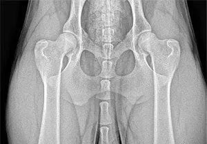
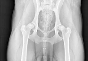
Excellent
This classification is assigned for superior conformation in comparison to other animals of the same age and breed. There is a deep seated ball (femoral head) which fits tightly into a well-formed socket (acetabulum) with minimal joint space. There is almost complete coverage of the socket over the ball.
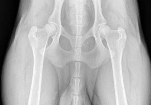
Good
Slightly less than superior but a well-formed congruent hip joint is visualized. The ball fits well into the socket and good coverage is present.
Fair
Assigned where minor irregularities in the hip joint exist. The hip joint is wider than a good hip phenotype. This is due to the ball slightly slipping out of the socket causing a minor degree of joint incongruency. There may also be slight inward deviation of the weight-bearing surface of the socket (dorsal acetabular rim) causing the socket to appear slightly shallow. This can be a normal finding in some breeds however, such as the Chinese Shar Pei, Chow Chow, and Poodle.
Borderline
There is no clear cut consensus between the radiologists to place the hip into a given category of normal or dysplastic. There is usually more incongruency present than what occurs in the minor amount found in a fair but there are no arthritic changes present that definitively diagnose the hip joint being dysplastic. There also may be a bony projection present on any of the areas of the hip anatomy illustrated above that can not accurately be assessed as being an abnormal arthritic change or as a normal anatomic variant for that individual dog. To increase the accuracy of a correct diagnosis, it is recommended to repeat the radiographs at a later date (usually 6 months). This allows the radiologist to compare the initial film with the most recent film over a given time period and assess for progressive arthritic changes that would be expected if the dog was truly dysplastic. Most dogs with this grade (over 50%) show no change in hip conformation over time and receive a normal hip rating; usually a fair hip phenotype.
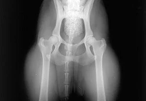
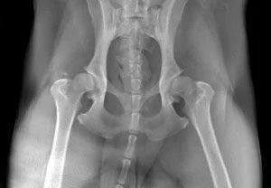
Mild
There is significant subluxation present where the ball is partially out of the socket causing an incongruent increased joint space. The socket is usually shallow only partially covering the ball. There are usually no arthritic changes present with this classification and if the dog is young (24 to 30 months of age), there is an option to resubmit an radiograph when the dog is older so it can be reevaluated a second time. Most dogs will remain dysplastic showing progression of the disease with early arthritic changes. Since HD is a chronic, progressive disease, the older the dog, the more accurate the diagnosis of HD (or lack of HD).
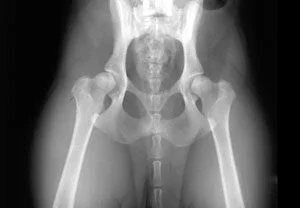
Moderate
There is significant subluxation present where the ball is barely seated into a shallow socket causing joint incongruency. There are secondary arthritic bone changes usually along the femoral neck and head (termed remodeling), acetabular rim changes (termed osteophytes or bone spurs) and various degrees of trabecular bone pattern changes called sclerosis. Once arthritis is reported, there is only continued progression of arthritis over time.
Severe
Assigned where radiographic evidence of marked dysplasia exists. There is significant subluxation present where the ball is partly or completely out of a shallow socket. Like moderate HD, there are also large amounts of secondary arthritic bone changes along the femoral neck and head, acetabular rim changes and large amounts of abnormal bone pattern changes.
The information above can be found on the OFA website.